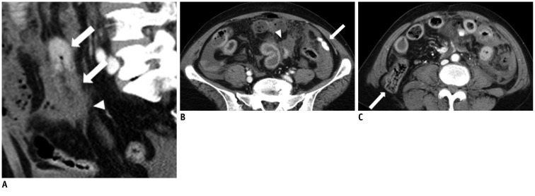
Fig. 4.
MDCT findings of multiple small bowel transections of 47-year-old woman.
A. Sagittal reformatted contrast enhanced multidetector CT scan shows complete cut off of bowel loop at proximal ileum. Ileal loop shows abnormal dual bowel wall enhancement (both increased and decreased); Janus sign (arrows). Surrounding hematoma (arrowhead) makes it difficult to recognize transected bowel end. B, C. Axial contrast enhanced multidetector CT scans show mesenteric extravasation (arrow in B), mesenteric hematoma (arrowhead in B) and lateral hernia of ascending colon (arrow in C).

