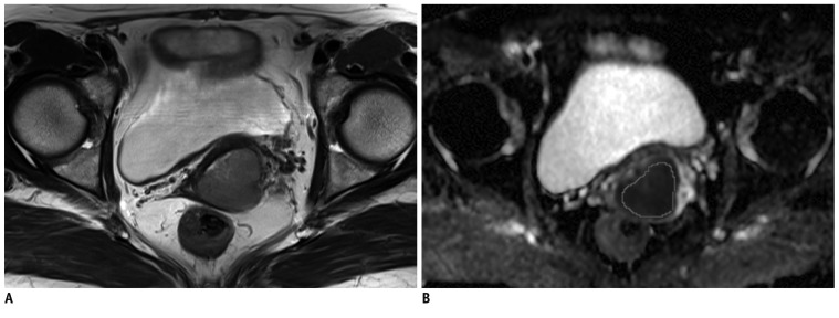
Fig. 1.
Method of measurement of ADC values in uterine cervical cancer.
Tumor border is identified by visual evaluation on axial T2-weighted image (A). ROI is manually drawn to include tumor as much as possible on ADC map (B) at level corresponding to A. ADC = apparent diffusion coefficient, ROI = region of interest

