Fig. 2.
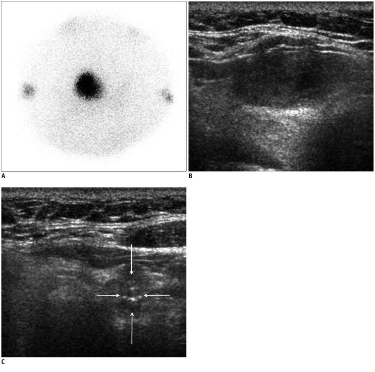
Hyperfunctioning cancer and coexisting cancer in 63-year-old woman (Patient No. 1).
A. Thyroid scan shows hot focus on right thyroid gland with suppressed surrounding normal tissue. B. On ultrasonography, low echoic, ovoid to round, smoothly marginated, solid nodule exists without any micro- or macro-calcification in right thyroid gland. It was categorized as indeterminate nodule of 2.6 cm maximal dimension, and could have been indicated with fine needle aspiration. It was surgically proven to be follicular thyroid cancer. C. On ultrasonography, low echoic, taller than wide, spiculated, solid nodule was observed with microcalcification in left thyroid gland (arrows). It was classified as suspicious malignant nodule of 1.1 cm maximal dimension, and could have been indicated for fine needle aspiration. It was surgically proven to be conventional papillary thyroid cancer.

