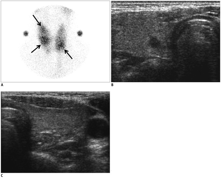
Fig. 3.
Coexisting cancer in 29-year-old woman (Patient No. 2).
A. On thyroid scan, 3 poorly defined hot foci were observed in both thyroid glands (arrows). B, C. On ultrasonography, no nodules which were correlated with thyroid scan could be seen at both thyroid glands. Instead, at right thyroid gland, low echoic, irregular, spiculated, solid nodule without any calcification was noted. It was classified as suspicious malignant nodule of 0.6 cm maximal dimension, and could have been indicated with fine needle aspiration. It was surgically proven to be conventional papillary thyroid cancer.

