Fig. 2.
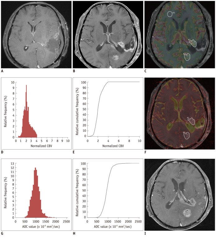
MR images, nCBV histograms and ADC histograms in 59-year-old man with glioblastoma.
A. Axial CE T1W image taken immediately after gross total resection shows no definite enhancing lesion. B. Three weeks after CCRT with TMZ, new enhancing lesions are visible in left periventricular white matter and in left occipital lobe. C. nCBV map, which is displayed as color overlay on CE T1W image taken 3 weeks after CCRT with TMZ, shows slightly increased nCBV in lesion (polygonal ROIs #1 and #2) compared with contralateral white matter (round ROI #3). D. Normalized CBV histograms and (E) cumulative histograms of enhancing lesions. F. ADC map, which is displayed as color overlay (in hot scale) on CE T1W image, shows slightly decreased ADC value for lesion (polygonal ROIs #1 and #2). G. ADC histograms and (H) cumulative histograms of enhancing lesion. I. After continuing TMZ for 1 month, patient visited emergency room due to involuntary movement. On second follow-up MR imaging that was performed during visit to emergency room, there was increase in enhancement of lesions without further treatment. After 3 months, patient passed away in spite of continuation of adjuvant TMZ, which is compatible with true progression. ADC = apparent diffusion coefficient, CBV = cerebral blood volume, CCRT = concurrent chemoradiotherapy, CE = contrast-enhanced, nCBV = normalized CBV, ROIs = regions of interest, TMZ = temozolomide, T1W = T1-weighted

