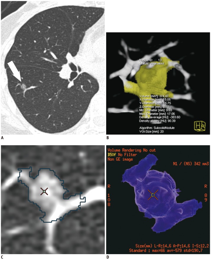
Fig. 3.
43-year-old woman with vessel-attached ground-glass nodule (GGN).
A. Transverse thin-section chest CT shows 7.3 mm pure GGN (arrow) in right lower lobe. B. Volume-rendered image of LungCARE shows poor segmentation of GGN with vascular segmentation leakage. C, D. LungVCAR provides segmentation boundary of GGN overlaid on transverse thin-section image (C) and volume-rendered image also shows poor segmentation of GGN due to attached vessels (D).

