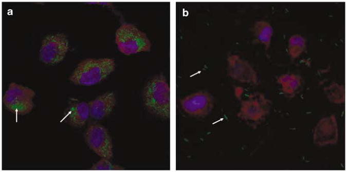
Figure 4.
Uptake of green fluorescent protein (GFP)-expressing invasive and non-invasive E. coli into cystic fibrosis tracheal epithelial (CFTE29o—) cells. CFTE29o— cells were infected with (a) invasive E. coli BM4570 (MOI 500) and (b) its non-invasive counterpart E. coli BM2710 (MOI 500) carrying the prokaryotic expression plasmid pAT505 (GFP expressed under the control of the prokaryotic Plac promoter). The cells were harvested 2 h post-infection. GFP-expressing bacteria appear in green, the cytosol and 4-6-diamidino-2-phenylindole (DAPI)-stained nuclei are shown red and blue, respectively. Original magnification × 63. Arrows indicate GFP-expressing E. coli.

