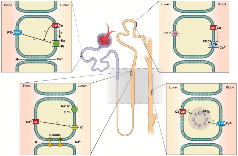
Figure 1.
Calcium reabsorption and calcium-sensing receptor in the nephron. (A) Proximal tubule: 50%–60% of filtered Ca2+ is reabsorbed paracellularly. Luminal calcium-sensing receptor (CSR) activation counteracts parathyroid hormone (PTH)-mediated Pi excretion, thereby promoting Pi conservation [11]. (B) Thick ascending limb: 30%–35% of filtered Ca2+ is reabsorbed paracellularly. Basolateral CSR inhibits potassium excretion via renal outer medullary potassium channel (ROMK), diminishing Ca2+ (as well as magnesium) reabsorption. Diminished potassium exit also reduces NaCl reabsorption via NKCl2, analogous to the effect of loop diuretics [12]. (C) Distal convoluted tubule: 10% of filtered Ca2+ is reabsorbed transcellularly. Basolateral CSR inhibits plasma membrane calcium ATPase (PMCA), thereby inhibiting transcellular Ca2+ reabsorption [10]. (D) Collecting duct: in the principle cells, activation of CSR inhibits retention of aquaporin-2 in the luminal-surface of the plasma membrane, thereby inhibiting antidiuretic hormone-mediated water conservation, causing renal water wasting [13,14]. In the intercalated cells, CSR promotes the activity of proton pump (H+-ATPase), enhancing urine acidification, thereby minimizing the risk of Ca2+ × Pi supersaturation [15].

