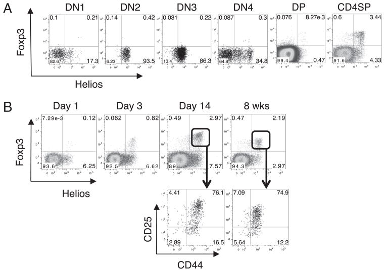
FIGURE 2.
Expression of Helios during thymic T cell development. A, Mouse thymocytes were stained for CD4, CD8, CD44, and CD25. DN, DP, and CD4SP cells were gated based on CD4 and CD8 expression and were further analyzed by flow cytometry for Helios and Foxp3. DN1–DN4 cells were gated on CD4−CD8− cells and further gated based on CD44 and CD25 expression. B, Thymocytes from two individual mice at the indicated ages were analyzed by flow cytometry for Helios and Foxp3 expression. Plots shown are gated on CD4+CD8− thymocytes. Mice were analyzed on different days but were always compared with an adult mouse. Shown are representative plots of one mouse from at least two independent experiments.

