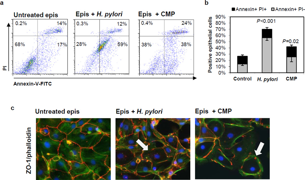
Figure 2. H. pylori induces apoptotic cell death in primary human gastric epithelial cells.
(a, b) Gastric epithelial cells were cultured for 3 days on collagen-coated plates and then treated with H. pylori (2×10⁷/mL), camptothecin (CMP, 5 µM) or medium alone. Apoptosis was determined by flow cytometric analysis of Annexin-V-FITC/propidiumiodide (PI)-stained cells after 6–8 h. (a) Representative data and (b) cumulative data from 10 (untreated, H. pylori) and 5 (CMP) experiments; mean ± SEM. (c) Microscopic analysis of ZO-1-Cy3/phalloidin-FITC/DAPI-stained epithelial monolayers after 24 h exposure to H. pylori or camptothecin. Arrows indicate areas with altered ZO-1 immunolocalization. Results are from a representative experiment (n=4).

