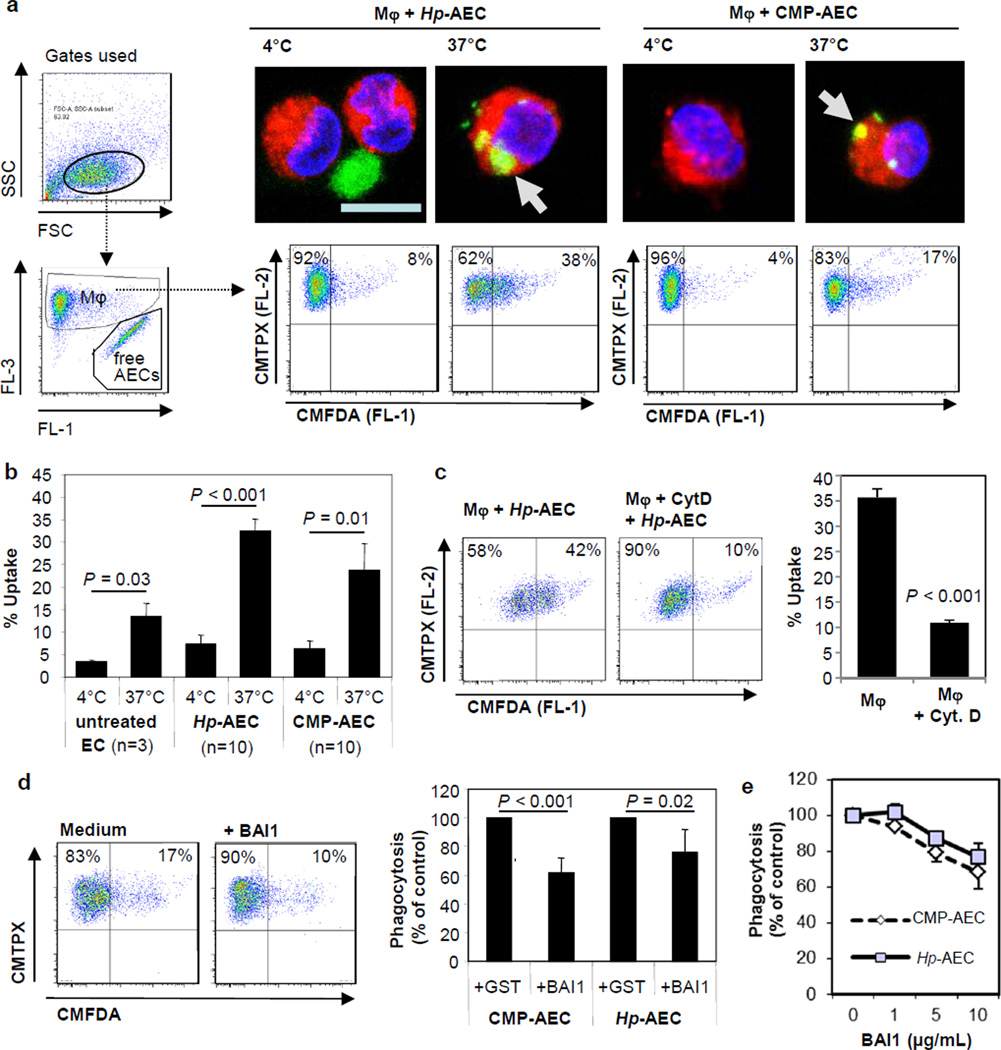
Figure 3. Apoptotic gastric epithelial cells are phagocytosed by monocyte-derived macrophages through a phosphatidylserin-dependent pathway.
(a, b) Gastric epithelial cell cultures were treated with H. pylori (Hp), camptothecin (CMP, 5 µM) or with medium alone for 6–8 h to induce apoptosis and then labeled with CMFDA (green). Monocyte-derived macrophages (MΦ) were stained with CMTPX (red), and equal numbers of stained macrophages and apoptotic epithelial cells (AECs) were co-cultured at 4°C or 37°C for 2.5 h. (a) Macrophages were analyzed by confocal microscopy (right, upper panels) or by flow cytometry (right, lower panels, bar = 10 µm). Representative data; panels on the left show gating strategy. (b) Cumulative data (mean ±SEM) from 3 (untreated) or 10 (Hp, CMP) experiments. (c) To block uptake, macrophages were pre-treated with cytochalasin D (1 µg/mL) for 45 min prior to co-culture with H. pylori-treated AECs (cytochalasin D also present during co-culture). Representative (left) and cumulative data (right), n=3; ***P ≤ 0.001. (d, e) Gastric epithelial cells with apoptosis induced by CMP (CMP-AEC) or H. pylori (Hp-AEC) were treated for 15 min with control GST or recombinant BAI1 RGD-TSR, which neutralizes surface phosphatidylserine, and then cultured with macrophages. (d) Representative (left) and cumulative data (right) from 4 experiments with 10 µg/mL of BAI1 RGD-TSR or control GST. Phagocytosis is expressed as % macrophages that contained BAI1-treated AECs relative to GST-treated AECs (100%). (e) GST and BAI1 used at the indicated concentration, mean ± SEM of 2 (camptothecin) or 3 (H. pylori) experiments.

