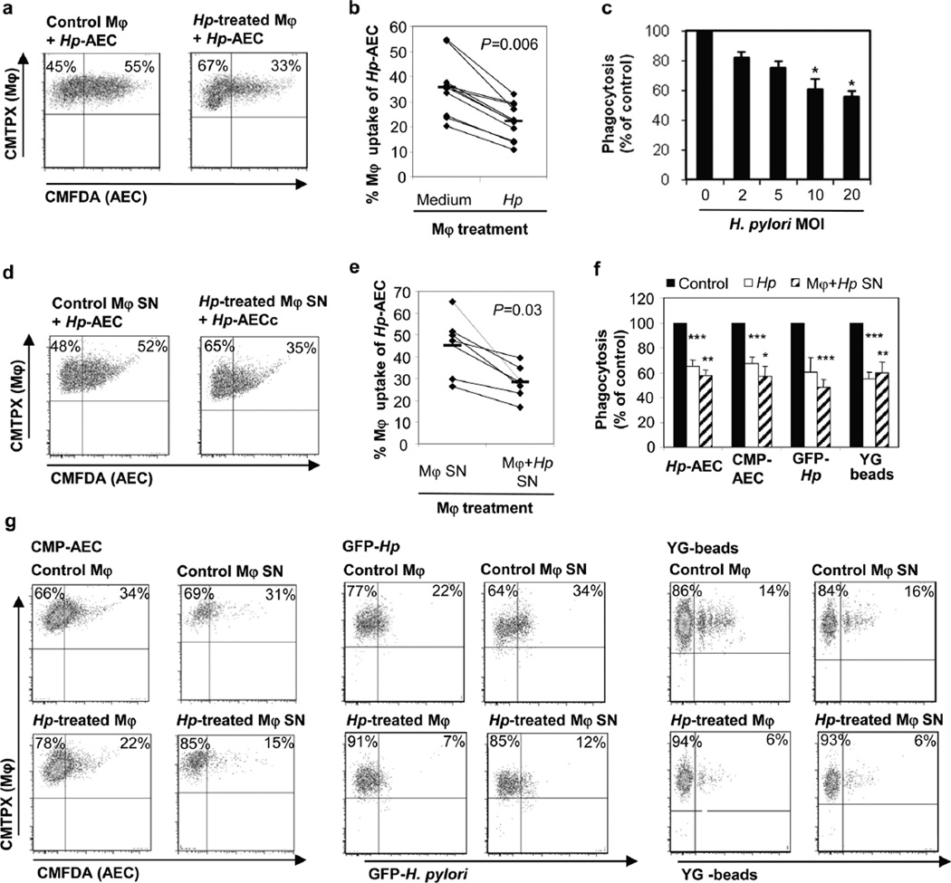
Figure 4. H. pylori stimulation inhibits macrophage phagocytic activity for apoptotic gastric epithelial cells.
(a, b) Monocyte-derived macrophages were treated with H. pylori (MOI=10) for 6–8 h or left untreated and then stained with CMTPX (red). Gastric epithelial cells were cultured for 3 days on collagen-coated plates, treated with H. pylori (2×10⁷/mL) or camptothecin (5 µM) for 6–8 h and labeled with CMFDA (green). Macrophages and apoptotic epithelial cells then were harvested and incubated at a ratio of 1:1 at 37°C for 2.5 h to allow macrophage engulfment of epithelial cells. (a) Representative FACS plots and (b) individual values (diamonds) and means (bars), n=10. (c) Phagocytosis of AECs after macrophage pre-treatment with different concentrations of live H. pylori, n=3, mean ± SEM, 1-way ANOVA with Tukey’s post hoc test * P≤0.05. (d, e) Soluble mediators released by H. pylori-treated macrophages inhibit AEC phagocytosis. Cell-free supernatants of macrophages cultured with or without H. pylori for 6–8 h were added to untreated macrophages for 6 h prior to the phagocytosis experiment. (d) Representative FACS plots and (e) individual values (diamonds) and means (bars), n=6. (f, g) H. pylori-induced inhibition of macrophage phagocytosis is not specific to the uptake of AECs. Macrophages were pre-treated with either H. pylori, culture supernatants from H. pylori-treated macrophages or media and then assayed for phagocytosis of Hp-AEC, CMP-AEC, GFP-labeled H. pylori (MOI=50) or YG fluorescent latex beads (40 beads/cell). (f) The relative efficiency of H. pylori-treated macrophages was determined by comparing the phagocytosis of treated macrophages to that of untreated macrophages for the different targets, mean ± SEM, n=4. (g) Representative FACS plots for the data shown in (f).

