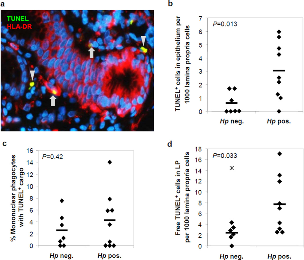
Figure 6. Increased epithelial cell apoptosis and decreased apoptotic cell clearance in H. pylori infected human gastric mucosa.
Paraffin-embedded gastric tissue from seven non-infected and nine H. pylori-infected human subjects was quantitatively analyzed for HLA-DR+ mononuclear phagocytes, TUNEL+ apoptotic cells/cell fragments and DAPI+ nuclei. (a) Gastric mucosa of an H. pylori-infected subject. Arrows: HLA-DR-Cy3+ (red) mononuclear phagocytes containing TUNEL-FITC+ (green) apoptotic material; arrowheads: non-phagocytosed TUNEL+ apoptotic material. Note that some glandular epithelial cells express HLA-DR. Original magnification 400x. (b) Frequency of TUNEL+ cells in gastric epithelial layer relative to total lamina propria cells. (c) Percentage of HLA-DR+ mononuclear phagocytes containing TUNEL+ material. (d) Free TUNEL+ material not associated with HLA-DR+ mononuclear phagocytes in gastric lamina propria relative to total lamina propria cells. Mean (bars) and individual values (diamonds) are shown; star – patient #3, outlier, excluded from analysis (value – 3 SD from mean); Student’s t test.

