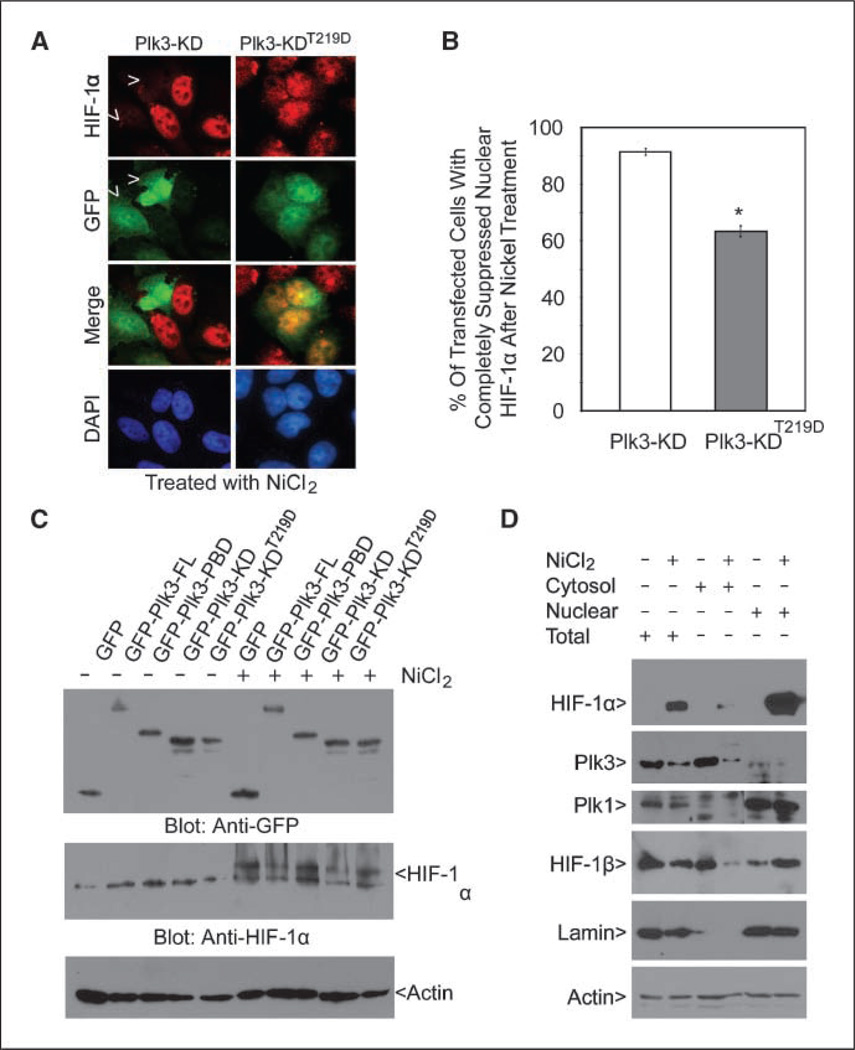
Figure 5.
HIF-1α expression is negatively correlated with Plk3 activity. A, HeLa cells transfected with GFP-Plk3-KD or GFP-Plk3-KDT219D expression constructs for 24 h and then treated with NiCl2 for 4 h were stained with antibodies to HIF-1α (red) and GFP (green). DNA was stained with DAPI (blue). Representative cells are shown. Arrows, Plk3-KD–expressing cells with suppressed accumulation of nuclear HIF-1α after NiCl2 treatment. B, HeLa cells transfected with GFP-Plk3-KD or GFP-Plk3-KDT219D expression constructs for 24 h and then treated with NiCl2 for 4 h were stained with antibodies to HIF-1α and GFP. The percent of transfected cells with completely suppressed nuclear HIF-1α after nickel ion treatment was summarized from three independent experiments. *, significant statistical difference (P < 0.01). C, HeLa cells were transfected with various GFP-Plk3 expression constructs as indicated for 24 h followed by treatment with NiCl2 for 4 h. Equal amounts of cell lysates were blotted for GFP, HIF-1α, and β-actin. D, HeLa cells were treated with or without NiCl2 for 24 h. Equal amounts of total cell lysates or cytoplasmic/nuclear fractions were blotted for HIF-1α, Plk3, Plk1, HIF-1β, lamin B, and β-actin. Lamin B, a nuclear protein, was examined to confirm effective subcellular fractionation. Each experiment was repeated at least thrice.

