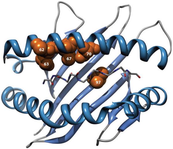
Figure 3.
Three-dimensional ribbon representation of the HLA-B protein showing amino acid positions 62, 63, 67, 70, and 97 lining the peptide-binding groove. The peptide backbone of the epitope is also displayed. Adapted from Reference 5 with permission.

