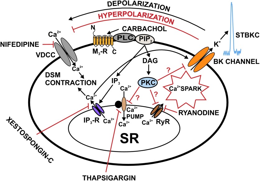
Fig. 8.
Schematic diagram illustrating the mechanisms by which M3-R regulates BK channel function in rat DSM cells. Activation of M3-Rs leads to IP3 and diacylglycerol (DAG) production, via a pathway involving PLC and PIP2. IP3 activates IP3-Rs, which releases Ca2+ from the SR. This IP3-induced Ca2+ release transiently activates the BK channels. Over time, depletion of SR Ca2+ upon activation of M3-Rs reduces Ca2+ spark activity, which leads to inhibition of STBKCs and depolarization of DSM cell RMP causing activation of VDCC, and thus increases DSM contractility. DAG activates PKC, which may lead to direct inhibition of the SR Ca2+ pump and RyRs resulting in suppression of Ca2+ sparks and STBKCs. Pharmacological tools used in this study to inhibit cellular sources of Ca2+ are indicated. DAG, diacylglycerol; PIP2, phosphatidylinositol 4,5-bisphosphate; PKC, protein kinase-C; PLC, phospholipase-C; VDCC, L-type voltage-dependent Ca2+ channel.

