
Fig. 5.
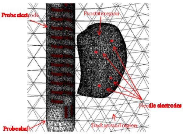
A reconstruction FE mesh with defined probe electrodes, prostate region, probe shaft region, background region and needle electrodes. The surface of a prostate was captured by TRUS in a patient and the locations of 4 simulated needle electrodes were indicated by red dots.