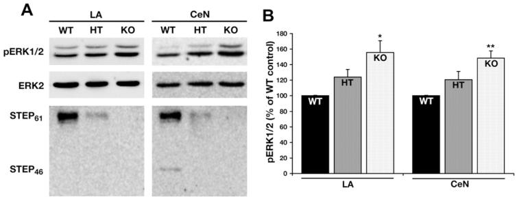
Fig. 2.
Increased amygdala ERK1/2 activity in STEP−/− mice. (A) Tissue punches from the LA and the CeN were analyzed by SDS–polyacrylamide gel electrophoresis and Western blots probed with anti-ERK1/2, anti-pERK1/2 and anti-STEP antibodies to determine the level of ERK1/2 activity and distribution of STEP isoforms in these regions. (B) Histograms showing an increase in pERK in both the LA and the CeN of STEP−/− mice (*p ≤ 0.05 and **p ≤ 0.01).

