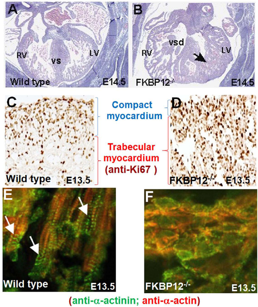
Figure 3.
Hypertrabeculation and noncompaction in FKBP12-deficient hearts. A and B, Cardiac histology of wild-type (A) and FKBP12-deficient heart (B) at E14.5. Black arrow denotes persistent trabecular myocardium in FKBP12 mutant heart. LV, left ventricle; RV, right ventricle; VS, ventricular septum; VSD, ventricular septal defect. C and D, Marked increase of cardiomyocyte proliferation in FKBP12 mutant heart. Immunohistochemical analysis of anti-Ki67 immune reactivity; the dark-brown nuclear signals are positive for Ki67, indicating proliferating cells. E and F, Disrupted cardiomyocyte polarization and myofibrillogenesis in FKBP12 mutant trabecular myocardium. Immunofluorescence staining using anti-α-actinin and anti-α-actin antibody, white arrows denote well organized sarcomeres in elongated normal trabecular cardiomyocytes.

