Fig. 3.
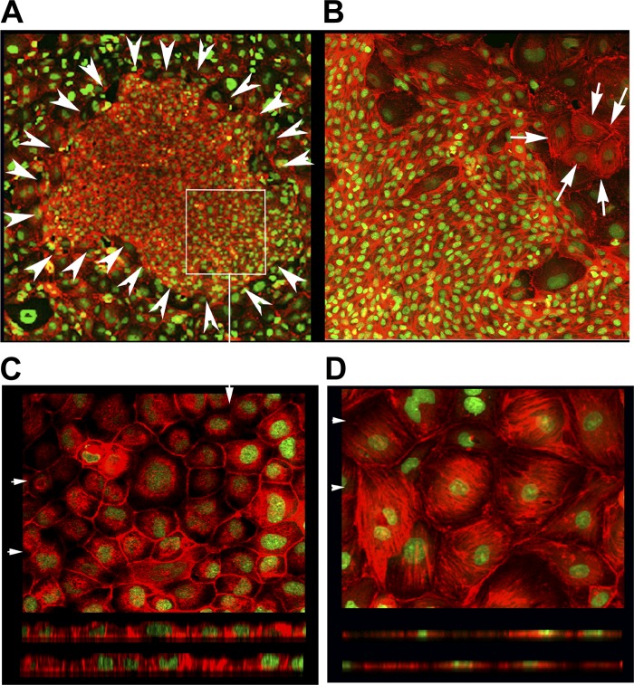
Typical F-actin staining results of clone C monolayers seeded at low density in the presence of 30 and 60 μg/ml galectin-3. A: clone C cells seeded at low density were cultured in the presence of 30 μg/ml recombinant rabbit galectin-3 for 4–5 days and stained with rhodamine phalloidin (F-actin, depicted in red) and SYTOX Green nuclear stain (×10 field). Arrowheads circumscribe a typical patch of HD phenotype cells that formed out of the LD monolayer in the presence of recombinant galectin-3 during culture. B: representative ×20 field of a LD clone C monolayer cultured in the presence of 60 μg/ml recombinant rabbit galectin-3 for 4 days shows the presence of a patch of HD phenotype cells adjacent to LD phenotype cells exhibiting stress fibers (the latter marked by arrows). C: high-magnification (×40) field of the patch area marked with a rectangular box in A. Top: XY projection of the top 4 optical sections (of 0.6 μm) revealing the presence of punctate apical actin. Bottom: actin staining (red) could be easily seen above the nucleus (apical portions) in the XZ projections taken at 2 places marked by arrowheads at top. D, top: typical XY projection of an LD monolayer field (×40) reveals the presence of exuberant stress fibers. Bottom: XZ sections of this field; note the absence of red (actin) staining above the nucleus.

