Fig. 2.
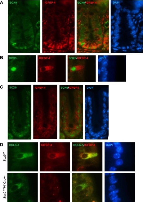
Representative images of immunofluorescence of SOX9 and IGFBP-4 of mouse intestinal crypts (A and C) and villi (B and D). A: higher expression of SOX9 (green) and IGFBP-4 (red) in Paneth cells and lower expression of IGFBP-4 and SOX9 in crypt base columnar cells. B: SOX9 (green) and IGFBP-4 (red) are colocalized in the solitary cells in the small intestinal epithelial villi. C: SOX9 (green) and IGFBP-4 (red) are also localized to the lower half of the crypts in mouse colons. D: IGFBP-4 (red) is observed in doublecortin-like kinase 1 (DCLK-1)-expressing tuft cells (green) in the wild-type mice (top), whereas IGFBP-4 is no longer expressed in DCLK-1-positive cells of Sox9-deficient mice (bottom).

