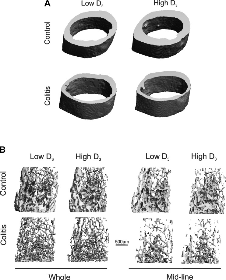
Fig. 5.
Trabecular and cortical bone morphology in control and IL-10−/− CD4+ T cell-transferred mice on the low or high vitamin D3 diet. Representative micro-CT (μCT) 3-dimensional reconstruction images show cortical bone of the femoral midshaft (A) and trabecular bone of the distal femoral metaphysis [B: complete thickness (whole) and midline (sagittal)].

