Figure 13.

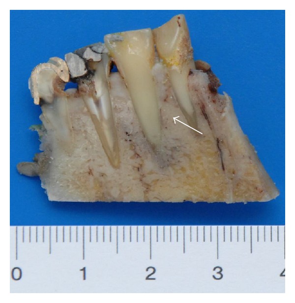
Case 4—the split resection specimen from a lingual view demonstrates the association of tumor formation along the root of the left lower canine (white arrows) as also further invasion of the cancellous bone.

Case 4—the split resection specimen from a lingual view demonstrates the association of tumor formation along the root of the left lower canine (white arrows) as also further invasion of the cancellous bone.