
Figure 3.
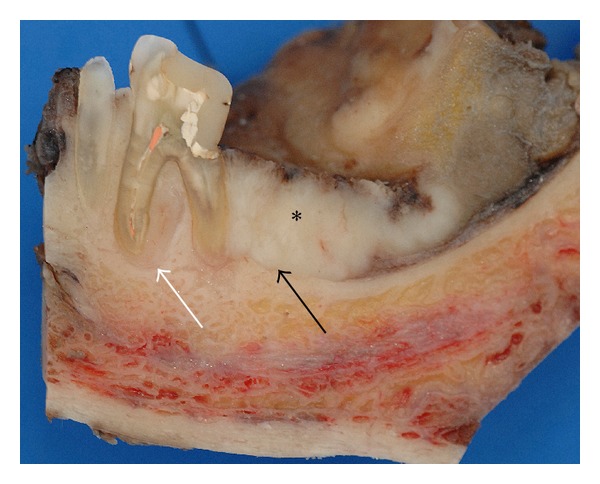
Case 1—cross section specimen. The black arrow highlights the close relation of the distal root to the tumor mass (∗) with preserved cortical bone distal to the tooth. The white arrow points at a tumor formation along the mesial root filling out the interradicular space.