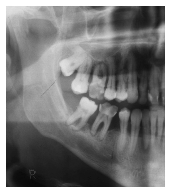
Figure 9.

Case 3—preoperative OPTG. Beside multiple carious lesions the present X-ray examination reveals signs of chronic periodontal disease with a general loss of horizontal bone level, liberation of both dental roots, and bifurcations. The local maximum of destruction is found in the region of the last two molars. The retromolar triangle, however, appears to be intact.
