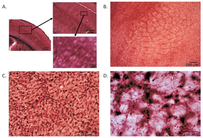
Figure 1.
P60 control tissue stained for myelin. Coronal section reveals myelinated axons in the barrel cortex running perpendicular to the pial surface terminating by layer IV (A). Low-magnification images reveal the barrel pattern (B), whereas higher magnifications (C and D) highlight individual myelinated axons cut in cross section in the tangential plane. Note that myelinated axons are sparser within the barrel hollows than in the septa/wall regions.

