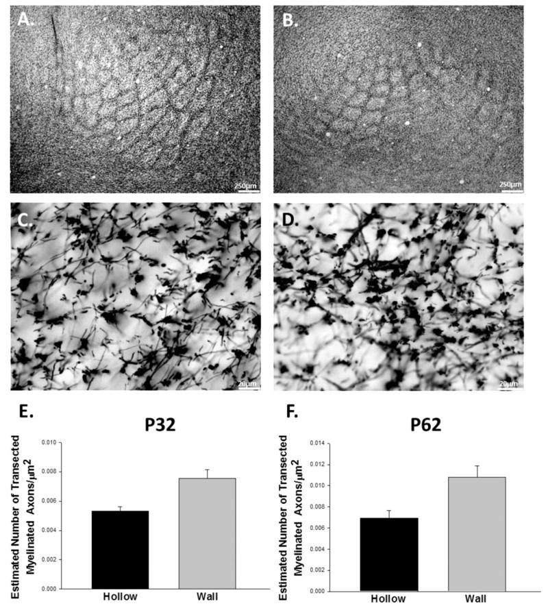
Figure 4.
Myelin basic protein staining in the mouse barrel cortex. Tangential sections at P32 tangential sections at low (A) and high magnifications (C) reveal a barrel pattern as do images from P62 animals at low (B) and high magnifications (D). Quantification of myelin basic protein + axons in the barrel hollow and barrel wall shows that barrel walls have greater densities of transected myelinated axons than barrel hollows at both time points (E and F).

