
Figure 1. Corpus Callosum Segmentation.
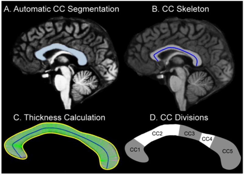
1A. Segmentation of the Corpus Callosum. The CC was identified in the transformed volume using an automated labeling procedure developed at the Martinos Center. 1B. CC Skeleton. A“skeleton” was generated by creating a line connecting the genu and the splenium such that the value of each voxel making up this line was equal to the minimum distance from the voxel to a point on the CC segmentation border. 1C. CC Thickness. The thickness at each of the 200 points along the skeleton was estimated as the distance between the superior and inferior borders with the constraint that the angle between these was greater than 3.0 radians. 1D.Topography of the midsaggital corpus callosum and proposed fiber composition, based on Hofer and Frahm’s classification. CC1 prefrontal, CC2 Premotor and supplementary motor, CC3-CC4 sensori-motor, CC5: parietal, temporal and occipital.