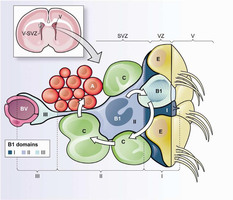
Figure 1. Schematic of the different domains of B1 cells within the adult V-SVZ.
Upper left. Frontal cross-section of the adult mouse brain showing the location of the ventricularsubventricular zone (V-SVZ), where neurogenesis in walls of the lateral ventricles (V) continues throughout life.
Lower panel. Cellular composition of the adult V-SVZ niche and domains of B1 cells. Neural stem cells (NSCs) correspond to type B1 cells (blue). B1 cells are surrounded by multiciliated ependymal cells (E) forming pinwheel-like structures on the ventricular surface. B1 cells give rise to intermediate progenitors (IPCs or C cells, green), which correspond to transit-amplifying cells that divide to generate neuroblasts (Type A cells, red). B1 cells retain epithelial properties, with a thin apical process (containing a primary cilium) that contacts the lateral ventricle (V), and a long basal process ending on blood vessels (BV, purple). Therefore, B1 cells can be subdivided into three domains. Domain I (proximal or apical, dark blue) contains the primary cilium and is in direct contact with the CSF; within this domain B1 cells can access soluble factors within the CSF and signaling molecules from neighboring ependymal cells. Domain II (intermediate, medium blue) is in close proximity to IPCs, neuroblasts, neuronal terminals and other supporting cells; cell-cell interactions between B1 cells and their progeny could occur within this compartment. Domain III (distal, light blue) comprises a basal process ending in a specialized end-foot that contacts blood vessels; blood-borne factors and endothelial-derived factors may act on B1 cells in this domain.

