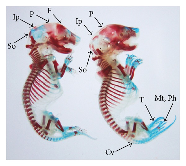
Figure 8.

Photograph showing skeleton of fetuses with reduced ossification of frontal (F), parietal (P), intraparietal (Ip), supraoccipital (So) skull bones, and caudal vertebrae (Cv). Absence of tarsals (T), metatarsals (Mt), and phalanges (Ph) at the dose of 92.25 mg Ni/kg b.wt. as NiCl2·6H2O.
