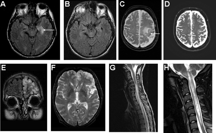
Figure 1.
Characteristic MRI brain and spinal cord findings. A, T2 FLAIR axial of brain demonstrating left hippocampal T2 abnormality typical of limbic encephalitis, which resolved with immunotherapy, B. C, T2 FLAIR axial image of brain demonstrating a superior–posterior left frontal abnormality in a patient who presented with right upper extremity apraxia and myoclonus. D, Postimmunotherapy improvement. E, T2 coronal FLAIR demonstrated extensive, predominantly left frontal cortical abnormality in a patient with ANNA-1 (anti-Hu), also seen on T2 axial image F. G, Sagittal T2 MRI of spinal cord demonstrates longitudinally extensive myelitis typical of NMO. H, Sagittal T2 MRI image of a patient with NMO who presented with intractable vomiting and myelitis. The longitudinally extensive lesion extends into the brain stem. Reproduced with permission from American Medical Association (C-F)20 and Lippincott Williams & Wilkins (H).36 MRI indicates magnetic resonance imaging; NMO, neuromyelitis optica; ANNA, antineuronal nuclear antibody.

