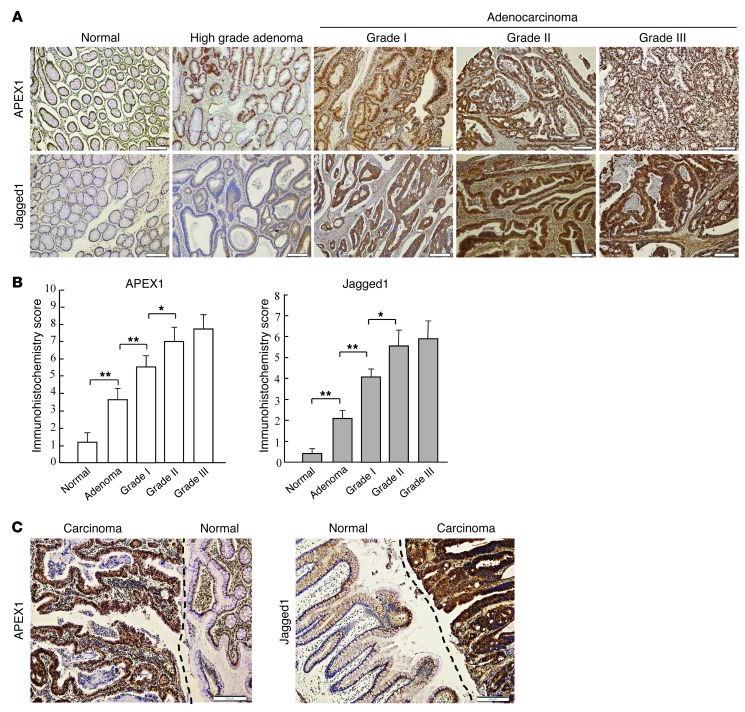
Figure 11. Correlation between APEX1 and Jagged1 expression in human colon cancer.
(A) Jagged1 and APEX1 proteins in normal colon tissue, colorectal adenoma, and grade I–III colorectal adenocarcinoma are shown by immunohistochemistry with anti-Jagged1 and anti-APEX1 antibodies. Brown staining indicates positive APEX1 or Jagged1 staining. (B) APEX1 and Jagged1 expression levels, assessed by immunohistochemistry scoring (see Methods). Jagged1 expression significantly correlated with APEX1 levels (P < 0.01, Pearson correlation test). (C) Representative images of Jagged1 and APEX1 immunoreactivity in normal colon epithelium and colon adenocarcinoma (separated by dashed lines). Results in B are SEM. *P < 0.05; **P < 0.01. Scale bars: 200 μm (A and C).

