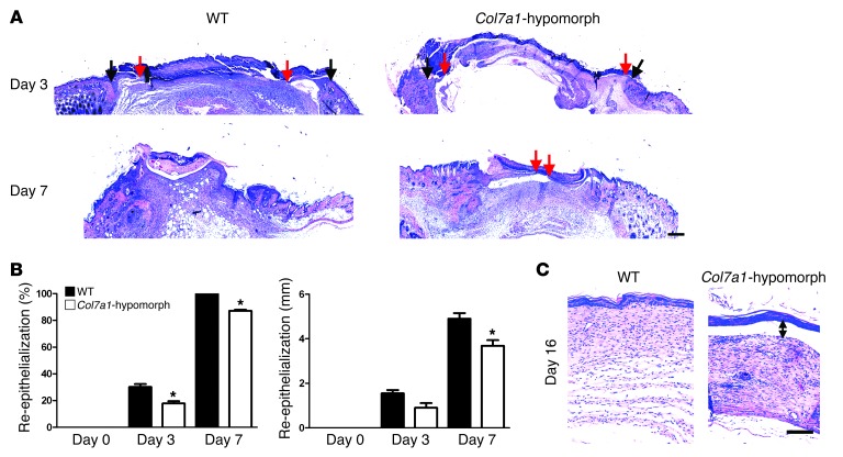
Figure 2. Loss of Col7a1 delays re-epithelialization.
(A) H&E staining of wounds in wild-type and Col7a1-hypomorphic mice at days 3, 7, and 16. Arrows indicate wound width (black arrows, initial wound border; red arrows, epithelial front). The original wound size was similar, but re-epithelialization was clearly protracted in Col7a1-hypomorphic wounds. Scale bar: 500 μm. (B) Quantification of percent re-epithelialization (left) and migration distance of the epithelial tongue (right). COL7A1 loss caused a significant delay in re-epithelialization. n ≥ 3 wounds; values represent mean ± SD. *P < 0.05. (C) Wounds after 16 days stained with H&E; note the detached epidermis in the Col7a1-hypomorphic wound (double-headed arrow). Note also the changes in the granulation tissue (see also Figure 6 and Supplemental Figure 12). Scale bar: 100 μm.

