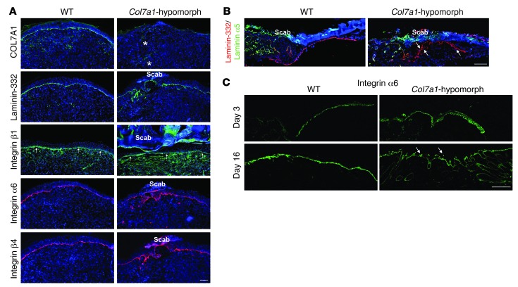
Figure 3. Loss of COL7A1 alters laminin-332 deposition and integrin α6β4 distribution in healing epidermis.
(A) 7-day wounds stained for laminin-332 and its integrin receptors. Laminin-332 deposition and integrin α6β4 distribution was altered in Col7a1-hypomorphic wounds. Asterisks show autofluorescence from red blood cells trapped in capillaries; arrows point to integrin β1 at the DEJZ. Scale bar: 100 μm. (B) Epidermal tongue in 3-day wounds stained for laminin-332 (red) and the laminin α5 chain (green). Laminin-332 deposition was irregular and patchy in Col7a1-hypomorphic wounds (arrows), in contrast to the distinct linear signal of the laminin α5 chain. Scale bar: 50 μm. (C) 3- and 16-day-old wounds stained for integrin α6. Arrows indicate the patchy suprabasal integrin α6 expression in 16-day-old Col7a1-hypomorpic wounds. Scale bar: 50 μm.

