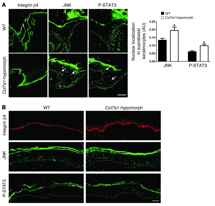
Figure 4. Abnormal activation of the laminin-332/integrin α6β4 signaling axis in Col7a1-hypomorphic wound epidermis.
(A) Epidermal tongue of 3-day-old wild-type and Col7a1-hypomorphic wounds stained for integrin β4, JNK, and phospho-STAT3. Note the abundant suprabasal presence of nuclear JNK and phospho-STAT3 in Col7a1-hypomorphic wounds (arrows), indicative of suprabasal activation. Quantification of staining is also shown (n ≥ 3; values represent mean ± SD). *P < 0.05. Scale bar: 50 μm. (B) Middle area of 7-day-old wild-type and Col7a1-hypomorphic wounds stained for integrin β4 (red), JNK (green), and phospho-STAT3 (green). Suprabasal activation persisted in older Col7a1-hypomorphic wounds. Scale bar: 50 μm.

