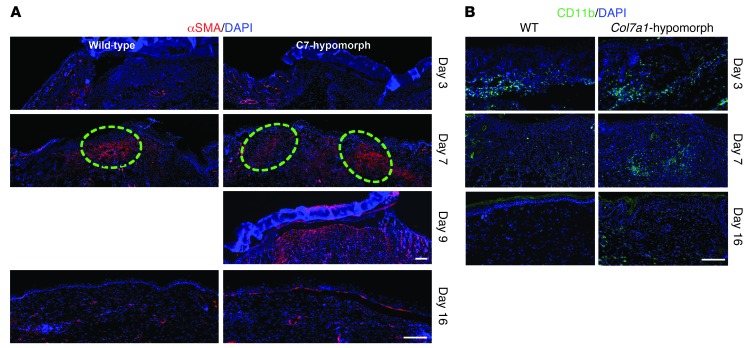
Figure 6. Granulation tissue maturation and clearance of inflammatory cells are delayed in Col7a1-hypomorphic wounds.
(A) α-SMA staining (red) showed few myofibroblasts at the wound edge at day 3 in both wild-type and Col7a1-hypomorphic mice. At day 7, myofibroblasts were abundant in the middle of the wild-type wound, but remained in the periphery of the Col7a1-hypomorphic wound. Dashed outlines denote the dense myofibroblast regions. Myofibroblast organization in the day 9 Col7a1-hypomorphic wound was similar to that of 7-day wild-type wounds, with myofibroblasts localized in the upper central part of the granulation tissue. At day 16 after wounding, most myofibroblasts had disappeared in both mice. Nuclei were stained with DAPI (blue). Scale bars: 100 μm. (B) CD11b staining (green) revealed protracted clearance of inflammatory cells in Col7a1-hypomophic wounds. At day 3, inflammatory cells were at the wound edge in both wild-type and Col7a1-hypomorphic mice. At day 7, inflammatory cells were mainly seen in the middle of the wound in both mice. At day 16, CD11b-positive cells were cleared from the wound area in wild-type mice, but not in Col7a1-hypomorphic mice. Nuclei were stained with DAPI (blue). Scale bar: 100 μm. See Supplemental Figure 12 for quantification of granulation tissue changes.

