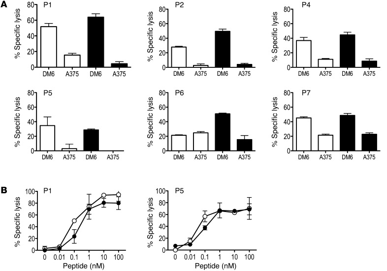
Figure 3. Vaccine-induced T cells recognize native gp100 and display high affinity for antigen.
(A) PBMCs collected after D3 were cultured in the presence of G209-2M (white bars and symbols) or G280-9V (black bars and symbols) peptide for 12 days as described in Figure 2 and tested in a standard 4-hour 51Cr release assay for their ability to recognize native gp100 antigen on human melanoma cells lines DM6 (HLA-A2+gp100+) and A375 (HLA-A2+gp100–). Percent specific lysis at a 30:1 effector/target ratio is shown; spontaneous lysis was <10%. Results are representative of 2 experiments. (B) Avidity of G209-2M– and G280-9V–specific T cells was determined in a standard 4-hour 51Cr release assay using peptide titrations and T2 (HLA-A*0201+) cells as targets. Percent specific lysis is shown for each peptide concentration; spontaneous lysis was <5%. Results are representative of 2 experiments.

