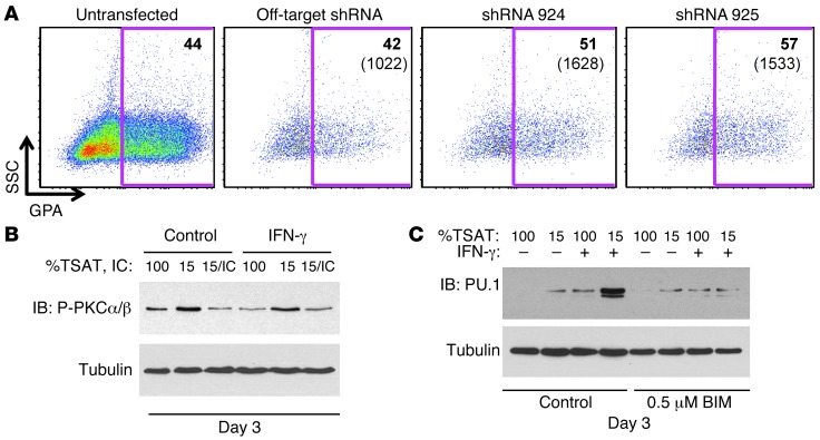
Figure 5. The cooperative induction of PU.1 by iron restriction and IFN-γ contributes to erythroid inhibition and requires PKC signaling.
(A) PU.1 knockdown enhances erythropoiesis in the setting of iron restriction plus IFN-γ stimulation. Human CD34+ cells were transduced with shRNA constructs, cultured 4 days in erythroid medium with iron restriction plus IFN-γ, and analyzed by flow cytometry with gating on GFP+ transduced cells. Relative percentage of GPA+ cells shown in top right corner; absolute number of GPA+ cells shown below in parentheses. Relative percentage of GFP+ cells and absolute number of GFP+ cells are as follows: off-target shRNA 27%, 3841; shRNA #924 35%, 4664; shRNA #925 39%, 3900. (B) Iron restriction induces PKCα/β hyperphosphorylation, IC reverses this effect, and IFN-γ shows no influence. Human CD34+ cells were cultured as in Figure 4A. (C) PKC signaling contributes to the cooperative induction of PU.1 by iron restriction and IFN-γ. Human CD34+ cells cultured as in Figure 4A were treated where indicated with 0.5 μM BIM, followed by immunoblot.

