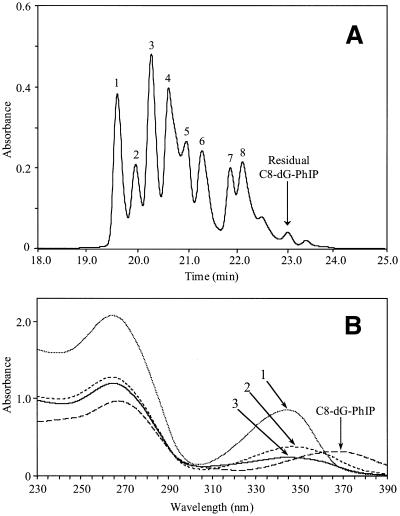
Figure 8.
(A) HPLC separation of the polar oligonucleotide adducts. Following large scale synthesis and isolation of the C8-dG-PhIP adduct, fractions containing the polar adducts were combined and separated on a semi-preparative column using a sodium phosphate to methanol gradient (0–27% over 17 min, isocratic for 13 min then to 100% over 5 min). Peaks 1, 2 and 3 were collected and purified by repeated HPLC. (B) Comparison of the UV spectra of oligonucleotide adduct peaks 1, 2 and 3 with the C8-dG-PhIP oligonucleotide adduct. Spectra were recorded in sodium phosphate (20mM, pH 7.0).

