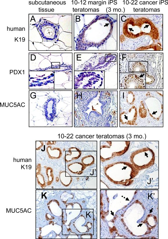
Figure 3. Teratomas at three months from 10-22 cancer iPS-like cells exhibit PanIN-like structures and marker expression.
A-C. No K19 staining in mouse subcutaneous tissue (negative control), weak K19 staining of 10-12 margin iPS-like teratomas at three months, and strong K19 staining of 10-22 cancer iPS-like teratomas at 3 months. D-I. Nuclear staining of PDX1 (arrow) and cytoplasmic staining of MUC5AC (arrow) only in teratomas of 10-22 cancer iPS-like teratomas at 3 months. J, K. Higher magnification shows uniform K19 staining (J, J’, arrow) and heterogeneous MUC5AC staining (K, K’) of teratomas from 10-22 cancer iPS-like cells.

