Figure 1. .
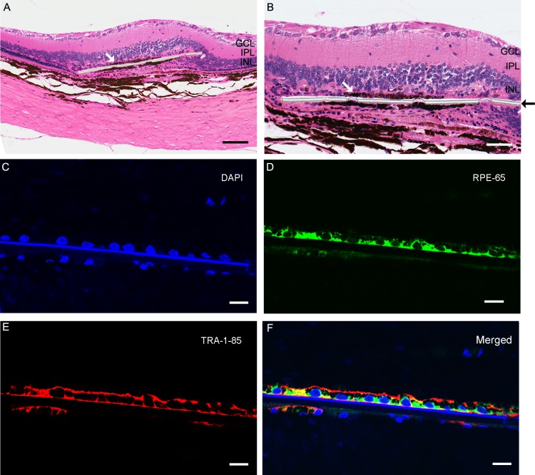
Implanted eye with parylene C membrane with cells 12 months after surgery. Monolayer of pigmented cells (white arrow) placed in the subretinal space over the parylene membrane (black arrow) observed with hematoxylin and eosin staining (A, B). Scale bars: A, 100 μm; B, 50 μm. Nuclei of implanted cells and host cells are counterstained by 4′,6-diamidino-2-phenylindole (DAPI) ([C], blue). Immunofluorescent staining for RPE65 ([D], green) and TRA-1-85 human marker ([E], red) was found in the transplanted cells. (F) Merged image of RPE65, TRA-1-85, and DAPI. Scale bars for C, D, E, F: 10 μm.

