
Fig 1.
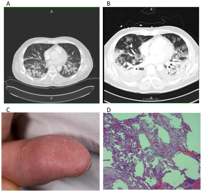
A) CT scan of chest with intravenous contrast 10 days before admission. The representative slice shows areas of patchy consolidation in the posterior portions of the lower lobes bilaterally. B) CT scan without intravenous contrast, taken on the day of admission, showing significant consolidation in the posterior aspects of the bilateral lower lobes, surrounded by patchy ground-glass infiltrates. C) Fingers of Case 1 showing dry, crackling skin at the fingertips, typical of mechanic’s hands. D) Hematoxylin and eosin stain of the left lingula showing areas of extensive interstitial fibroplasia and marked pneumocyte atypia and interstitial inflammation consistent with organizing diffuse alveolar damage with associated bronchopneumonia.