Figure 5.
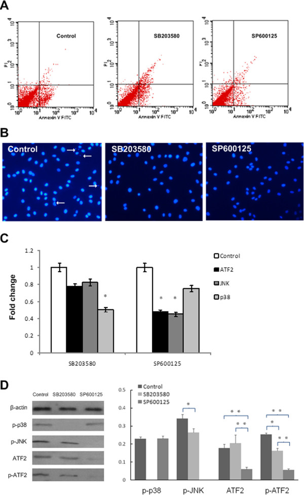
Analysis results of different treated groups. A. Apoptosis analysis of chondrocytes after different treatments for 3 days (control cultures with early apoptosis rate of 7.2 ± 1.3%; for cultures supplemented with p38 inhibitor SB203580 the rate was 5.8 ± 1.4%; those supplemented with JNK inhibitor SP600125 was 3.4 ± 1.1%). B. DAPI stain images of KBD chondrocytes in different groups (200 ×, control, SB203580 and SP600125, control groups showed the nuclear fragmentation and condensation). C. qPCR of ATF2, JNK & p38 in chondrocytes of different treatment normalized to GAPDH and normalized to control. SP600125 and SB203580 decreased the fold changes of ATF2 to 0.5 and 0.8 respectively, p < 0.001. D. Western blotting of p-p38, p-JNK, ATF2 and p-ATF2 in chondrocytes with different treatment. (The levels of p-p38, p-JNK, ATF2 and p-ATF2 were lower in chondrocytes cultured with inhibitors, and p-ATF2 was more likely to be influenced by JNK inhibitor). * p < 0.05, * * p < 0.01.

