
Figure 2. Lung tissue histopathology in TLR4-WT and TLR4-mutant mice following VILI.
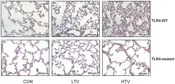
Representative photomicrographs of hematoxylin and eosin staining (× 20 objective) of lungs from TLR4-WT (A, B, C) and TLR4-mutant (D, E, F) mice subjected to unventilated and tracheotomized control (CON), low tidal volume ventilation (LTV), and high tidal volume ventilation (HTV). HTV illustrates a marked increase in inflammatory cell infiltration, alveolar septal thickening, and pulmonary edema in the lung in TLR4-WT mice (C) compared to TLR4-mutant mice (F).