Figure.

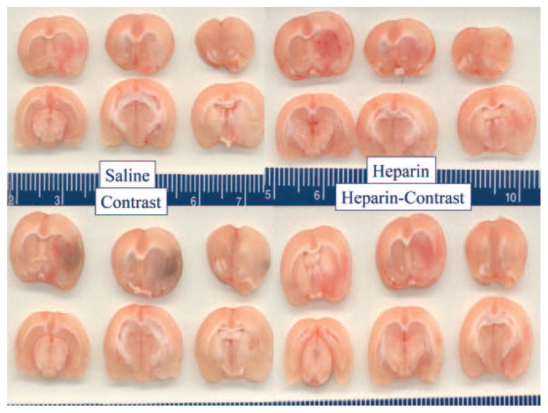
Postmortem brain sections from animals in the saline (top left), intravenous heparin (top right), contrast (lower left), and heparin+contrast (lower right) groups. Petechial hemorrhagic staining is visible in deep subcortical regions of all rats as well as in lateral cortical regions (reader’s right) of all animals. The blue color in the contrast group indicates greater petechial hemorrhagic change, including cortical regions.
