Figure 5.
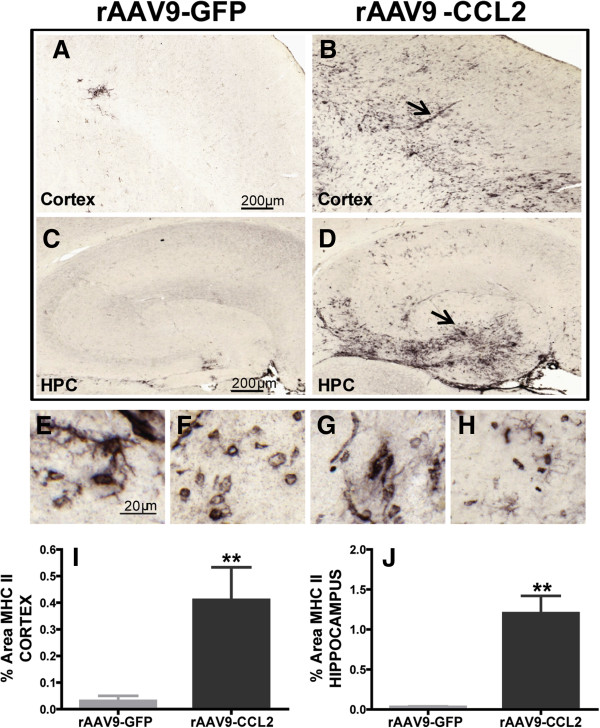
MHCII activation is induced in the brains of CCL2-overexpressing mice. (A) through (D) Immunohistochemical staining for MHCII showed that mononuclear cells/macrophages significantly increased MHCII expression following CCL2 overexpression. Images were collected from the cortices and hippocampi of wild-type mice injected with either rAAV9-GFP (A) and (C) or rAAV9-CCL2 (B) and (D). MHCII+ cells displayed various phenotypic morphologies, from rounded to ramified (arrow) and amoeboid (arrowhead) (E) through (H). Mean ± SEM percentage area for immunostaining of MHCII+ microglia in cortex and hippocampus are presented in (I) and (J). Student's t-test. (**P < 0.01, n = 6). Scale bars represent 200 μm and 20 μm.

