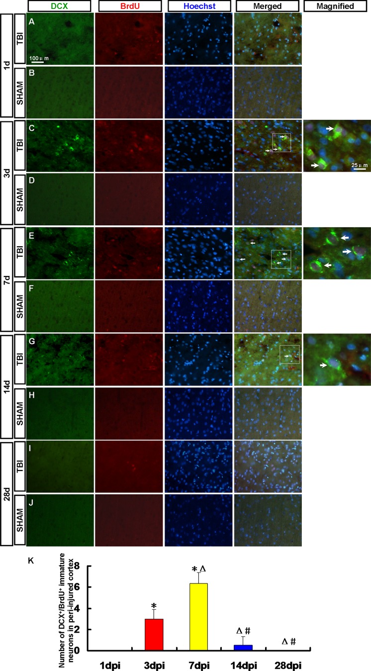
Figure 5. DCX+/BrdU+ newborn immature neurons in the peri-injured cortex.
Newborn immature neurons in the peri-injured cortex were detected by DCX (green) and BrdU (red) antibody. Arrows denote the DCX+/BrdU+ cells. (A, C, E, G, I) DCX+/BrdU+ immature neurons were observed in the peri-injured cortex at 1, 3, 7, 14 and 28 dpi. (B, D, F, H, J) DCX+/BrdU+ immature neurons were not found in the sham group. (K) A statistic diagram for the number of DCX+/BrdU+ cells in the peri-injured cortex at 1, 3, 7, 14 and 28 dpi. * p<0.05, vs. 1 dpi group; △ p<0.05, vs. 3 dpi group; # p<0.05 vs. 7 dpi group.

