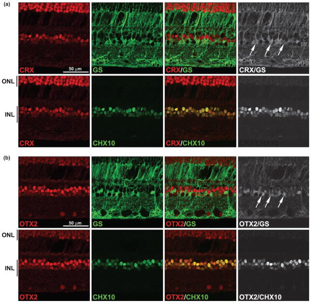
Fig. 5.
Co-immunostaining of CRX and OTX2 with markers of bipolar and Müller glial cells. A tissue section from an unaffected region of the eye of patient 35917 was triple-stained with rabbit anti-CRX antibody (a) or rabbit anti-OTX2 antibody (b), mouse anti-glutamine synthetase (GS) antibody and sheep anti-CHX10 antibody. Signals were detected with Alexa 555-conjugated donkey anti-rabbit (CRX, OTX2), Alexa 647-conjugated donkey anti-mouse (GS) and Alexa 488-conjugated donkey anti-sheep secondary antibodies (CHX10). Merging the CRX (a) or OTX2 (b) (red) and CHX10 (green) signals demonstrates their co-expression in bipolar cells (indicated by the yellow color). Merging the CRX (a) or OTX2 (b) (red) and GS (green) signals demonstrates their co-expression in Müller glial cells. Non-co-localized signals have been removed in the panels on the right in order to highlight sites of co-expression. Arrows point to CRX/GSand OTX2/GS-positive cells. INL, inner nuclear layer; ONL, outer nuclear layer (photoreceptor).

