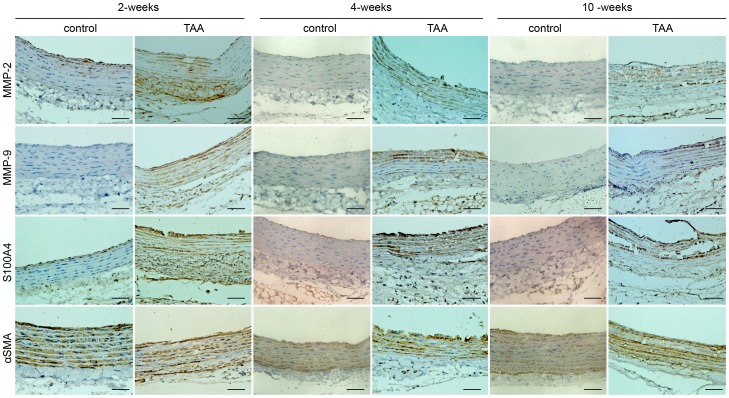
Figure 4. Representative pictures of immunohistochemical staining in aortic slides over time post-TAA induction.
The slides were stained with antibodies against MMP-2, MMP-9, S100A4 and αSMA. An anti rabbit HRP/DAB detection system was used to visualize expression (brown staining). Scale bar = 50 µm.

