Figure 6.

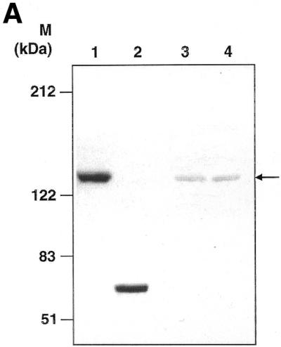
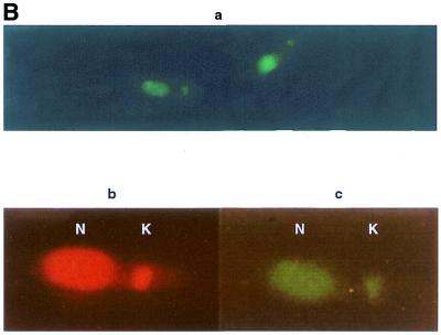
Western blotting and immunolocalisation studies using anti-GST–LdTOP2 antibody. (A) Western blot analysis of bacterial extract of induced pET3a–LdTOP2 (lane 1), purified GST–LdTOP2 fusion protein (lane 2), a leishmanial promastigote cell extract (lane 3) and a leishmanial amastigote cell extract (lane 4) reacted with the anti-LdTOP2 antibody. Arrow shows position of the L.donovani topoisomerase II. The standards for prestained broad range protein molecular weight marker (Bio-Rad) are shown on the left. (B) Immunocytochemical localisation with anti-LdTOP2 antiserum using fluorescent detection methods. Late log phase L.donovani promastigotes were fixed, and treated with the anti-LdTOP2 primary antibody and FITC-tagged secondary antibody. Parasite cells were also stained with ethidium bromide to locate the nucleus and the kinetoplast. (a) The visualisation of FITC fluorescence seen at 100× magnification in an Olympus fluorescence microscope. An enlarged view of a single promastigote after FITC staining (c) was obtained on scanning and it was compared with an ethidium bromide stained cell (b). The nucleus (N) and kinetoplast (K) are marked in b and c.

